DREAM OCT
Exploring the Whole Eye
- Deep
- Rapid
- Extensive
- Accurate
- Multimodal
ALL IN ONE
Technology
Deep
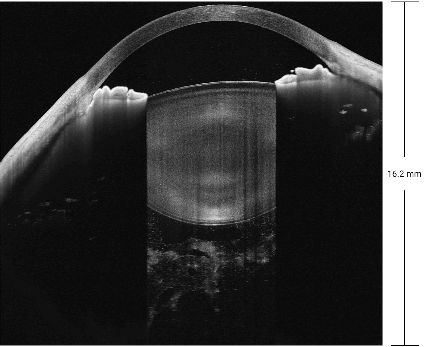
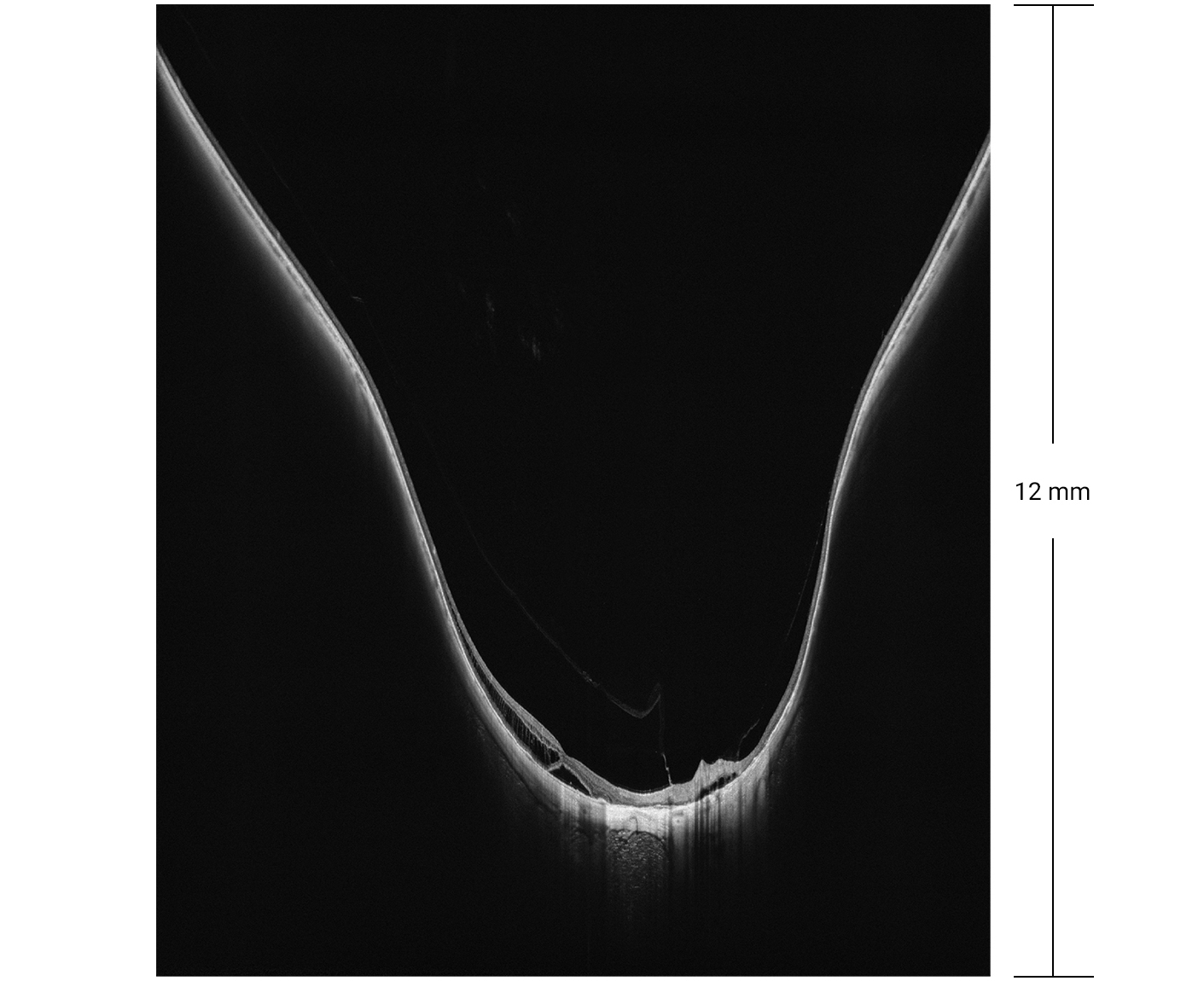
Based on Swept-Source OCT and an proprietary system, DREAM OCT™ can achieve an unprecedented imaging depth of 12 mm (in tissue) for retina imaging and 16.2 mm (in air) for anterior segment.
Rapid
With a scan speed up to 400KHz, DREAM OCT™ is faster than ever before. The high speed is crucial in high resolution OCT Angiography and eliminate the artefact caused by eye motions.
Comparison of Scan Speed
Extensive
DREAM OCT™ is able to cover an ultrawide field of 26 mm × 21mm (inner angle 130°) on retina with a single scan. An automatic montage can further extend the range to 200°, going beyond the entire posterior hemisphere and uncovering lesions toward the edge of retina.
Accurate
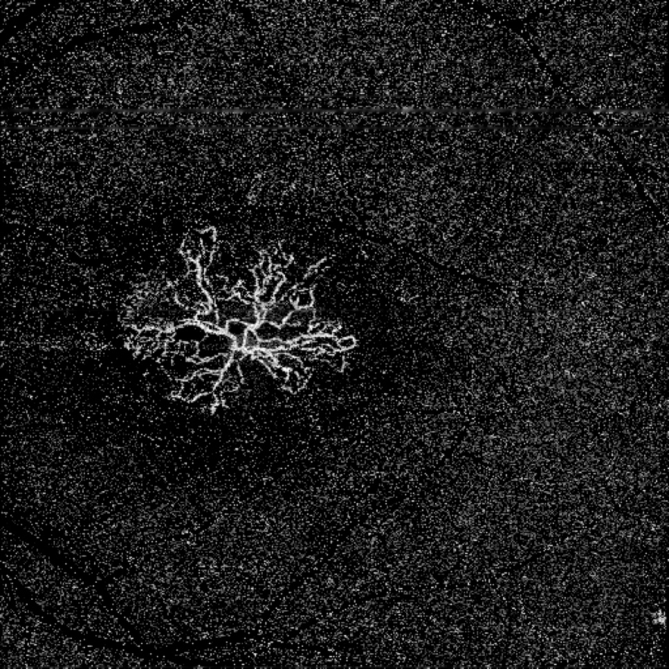
DREAM OCT™ is capable of detecting tiny nascent vascular pathologies while maintaining a low false positive rate. The proprietary algorithm of TrueAngio™ enables a high detection sensitivity of subtle flow signals. 3D Projection Artefact Removal and AI-based Deep Layer™ algorithms further help to reduce artefacts to reveal true lesions. In Anterior Segment imaging and biometric measurement, the raw images undergo sophisticated corrections to account for optical distortion and light refraction. It ensures an accurate and precise quantification of biometric parameters which are crucial for clinical decisions.


Multimodal
Powered by a state-of-the-art Swept-Source OCT engine, the DREAM OCT™ integrates a full set of imaging modalities including Retinal OCT, Retinal OCTA, AS-OCT, AS-OCTA, and visualized Biometry for the most challenging clinical and research applications. The combination of different modalities does not have to compromise each modality’s individual performance. Instead, they work together to collaboratively achieve functions that were impossible before, such as accurate measurement of retina curvature and full eye imaging that reconstructs geometrically accurate 3D representation of eyes.
Gallery


 & Choroidal Vessels Index (CVI) Analysis")


Literature
Appearance of Tumor Vessels in Patients With Choroidal Osteoma Using Swept-Source Optical Coherence Tomographic Angiography
Dietary ω-3 polyunsaturated fatty acids are protective for myopia
Miaozhen Pan, Fei Zhao, Bintao Xie, Hao Wu, Sen Zhang, Cong YeZhenqi Guan, Lin Kang, Yuqing Zhang, Xuan Zhao, Yi Lei, Qi Wang, Li Wang, Fan Yang, Chenchen Zhao, Jia Qu, Xi-angtian Zhou.
Dietary ω-3 polyunsaturated fatty acids are protective for myopia.
Proc Natl Acad Sci U S A, 2021 Oct 26.
doi: 10.1073/pnas.2104689118.
About
SVision Imaging, Ltd was founded by a group of scientists and industry veterans of Silicon Valley in 2014 with a core mission to develop the most advanced ophthalmic technologies. In 2015, the company’s base of operations moved to mainland China, and there are now three sites in Silicon Valley, Shanghai, and Luoyang.
In 2019, SVision released the first Swept-Source Optical Coherence Tomography (OCT) device that combines Deep imaging depth, Rapid sweeping speed, Extensive scan range, Accurate lesion detection and Multimodal imaging capabilities, abbreviated as DREAM OCT™.
The powerful imaging device has become an indispensable tool for many ophthalmologists in not only their daily clinical use but also their exploration of the research frontiers. To date, 47 papers have been published in peer-reviewed journals based on the findings using DREAM OCT™ devices.
SVision will continue the innovation and push the limit of technologies. The product portfolios are extending from ophthalmic diagnostic devices to operational instruments. In doing so, SVision aims to empower ophthalmologists and benefit the patients worldwide.
* The biometry module and the A-scan rate of 400 kHz has NOT got the FDA approval yet.
Silicon Valley Office: 160 E Tasman Drive, Suite 107, San Jose, CA 95134, USA
Shanghai Office: 3F, 939 Haining Road, Shanghai, 200085, China
Luoyang Office: 2 Penglai Road, Bldg 3A-1F, National University Science and Technology Park, Luoyang, Henan 471003, China
+86 400-0379-805
info@svr.us